Week Four Quiz
The quiz is divided into two sections. The first section contains questions that assess your recall of essential biological facts. The second set of questions asks you to apply your knowledge of material presented to solve clinical or research problems. The questions in the second set are similar to what you will encounter on the self-assessment and qualifier.
Instructions: To check your answer, click on the option you think is correct.
Application Questions
-
A 58-year-old woman has metastatic breast cancer. Her tumor cells express high levels of cyclin D. She starts palbociclib, a selective inhibitor of Cdk4/6, and her disease is stable for 14 months. Then new lesions appear. A biopsy of a new lesion shows the same cyclin D overexpression plus a new mutation in one gene. A loss-of-function mutation in which of the following genes would most likely explain why the drug stopped working?
- CDKN2A (encoding Ink4)
- MYC
- RB1 (encoding pRb)
- KRAS (encoding Ras)
- CCND1 (encoding cyclin D)
Show Explanation
-
A 64-year-old man has metastatic colorectal cancer that overexpresses EGFR, a receptor tyrosine kinase. His oncologist is thinking about cetuximab, an antibody that blocks EGF from binding EGFR. Molecular testing finds an activating mutation in codon 12 of KRAS, and the oncologist decides the drug will not help. What is the best explanation for this decision?
- The mutant Ras protein binds cetuximab and neutralizes it.
- The mutant Ras stays bound to GTP and keeps the MAP kinase pathway, Myc and cyclin D expression active without any signal from the receptor.
- The mutant Ras activates the TGF-β receptor, which blocks Smad signaling.
- The mutant Ras turns on p53, which makes the cells resistant to apoptosis.
- The mutant Ras stops EGFR from being made, so the drug has no target.
Show Explanation
-
A research laboratory creates a mouse model with knockout of the gene encoding superoxide dismutase 1 (SOD1). Compared to wild-type mice, these knockout mice are expected to die early from ROS-mediated disease. Which of the following best describes the normal function of SOD1 and the consequence of its absence?
- SOD1 converts H2O2 to water; its absence allows H2O2 to accumulate and cause DNA strand breaks
- SOD1 converts superoxide anion (O2-) to H2O2; its absence causes superoxide accumulation and widespread oxidative damage
- SOD1 repairs 8-oxo-G DNA lesions; its absence leads to unrepaired mutations and early-onset cancer
- SOD1 activates catalase; its absence impairs the breakdown of hydroxyl radicals
- SOD1 refolds oxidized proteins; its absence leads to protein aggregation and neurodegeneration
Show Explanation
-
A 52-year-old man with a history of alcoholic cirrhosis is brought to the emergency department after ingesting a large quantity of acetaminophen in a suicide attempt. Lab work reveals markedly elevated AST and ALT. A liver biopsy is performed and shows hepatocyte swelling, membrane disruption, and leakage of intracellular contents into surrounding tissue. On further workup, it is found that toxic metabolites of acetaminophen depleted hepatocellular glutathione stores. Which of the following best explains the mechanism by which glutathione depletion leads to hepatocyte death?
- Decreased ATP production due to inhibition of the mitochondrial electron transport chain
- Direct inhibition of superoxide dismutase, causing superoxide anion accumulation
- Activation of HSP70 chaperones leading to ubiquitin-proteasome degradation of hepatocytes
- Loss of antioxidant defense allowing ROS accumulation, leading to lipid peroxidation and membrane disruption
- Impaired DNA repair via inhibition of XPA-mediated nucleotide excision repair
Show Explanation
-
A 58-year-old man with a 30-year history of smoking presents for a routine chest CT, which reveals thickened bronchial walls. Bronchoscopic biopsy shows replacement of the normal pseudostratified columnar epithelium with stratified squamous epithelium. There are no nuclear atypia or architectural disturbances. Which of the following best describes the underlying mechanism driving this change?
- Somatic mutations in tumor suppressor genes
- Activation of proto-oncogenes by carcinogens
- Epigenetic changes including DNA methylation and gene silencing
- Mitotic spindle dysfunction leading to aneuploidy
- Increased ROS production causing direct nuclear damage
Show Explanation
-
A 72-year-old woman is diagnosed with severe aortic stenosis. Echocardiography shows markedly increased left ventricular wall thickness with preserved chamber size. Cardiac catheterization reveals elevated intraventricular pressures. Which of the following best describes the cellular changes occurring in the myocardium, and what is the expected long-term consequence if left untreated?
- Hyperplasia of cardiomyocytes; risk of arrhythmia
- Hypertrophy of cardiomyocytes; eventual heart failure and fibrosis
- Metaplasia of cardiomyocytes to fibroblasts; progressive wall thinning
- Atrophy of cardiomyocytes due to pressure overload; ventricular dilation
- Dysplasia of cardiomyocytes; progression to cardiac malignancy
Show Explanation
-
A 35-year-old woman is undergoing chemotherapy for breast cancer. Her oncologist explains that the drug works by causing DNA damage in rapidly dividing cells, triggering the intrinsic apoptotic pathway. Which of the following correctly describes the sequence of events in this pathway?
- FasL binds Fas → FADD recruitment → Caspase-8 activation → Caspase-3 activation
- Perforin pore formation → Granzyme entry → direct caspase activation
- Inflammasome activation → Gasdermin pore formation → IL-1β release
- Caspase-8 inhibition → RIPK3 activation → membrane rupture
- DNA damage → BAX/BAK activation → Cytochrome c release → Apoptosome formation → Caspase-3 activation
Show Explanation
-
A 52-year-old man is brought to the emergency department with severe epigastric pain that goes through to his back, plus nausea and repeated vomiting for 18 hours. He has drunk about 12 beers a day for 20 years. His temperature is 38.3°C (101°F), pulse 124/min, and blood pressure 88/54 mm Hg. The upper abdomen is tender with guarding, and there is bruising around the belly button. Lab results:
- Serum lipase: 2,850 U/L
- Serum calcium: 7.4 mg/dL
- Serum albumin: 4.0 g/dL
Despite aggressive fluids, he develops acute respiratory distress syndrome (ARDS) and dies on hospital day 3. A photo of his pancreas and surrounding tissue from the autopsy is shown.
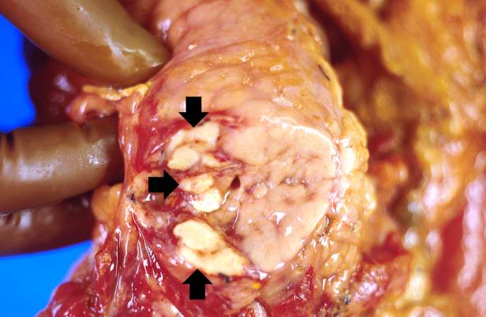
Which of the following processes best explains the chalky white lesions shown in the image?
- Protein denaturation from loss of blood supply, with the tissue's outline preserved
- Calcium deposits in normal tissue caused by high blood calcium
- Release of fatty acids by digestive enzymes, which then bind calcium
- Digestion of dead tissue by enzymes from white blood cells, leaving a liquid-filled cavity
- A cheese-like, crumbly mass of dead tissue inside a granuloma
Show Explanation