Cardiac Radiology
Learning Objectives
- Overview the currently available imaging modalities to evaluate the cardiac structures
- Review their strengths and weaknesses
- Review common indications for each modality
Overview
There are several imaging modalities that can evaluate the cardiac structures including chest radiography, echocardiography, computed tomography (CT), magnetic resonance imaging (MRI), Myocardial perfusion testing with Nuclear Medicine, and direct cardiac catheterization.
These modalities are usually complimentary and chosen on a given clinical scenario based one their strengths and weaknesses. Other factors that play a role while deciding which modality to choose from are cost, radiation exposure, test availability, required personnel training, length of examination, necessary patient cooperation, and the provided data in the acquired images.
Strengths and weaknesses
Each modality has its inherit strengths and weaknesses based on the how the images are acquired and the information they display.
Plain Radiography is a relatively affordable examination that is readily available in a hospital setting. Images are acquired fast. It exposes the patient to a relatively small amount of radiation but no intravenous contrast. It does not require extensive technologist training and it does not demand significant patient cooperation.
On the other hand, the images provided by radiography technique have low spatial resolution. A 3D volume is depicted as a 2D picture leading to superimposition of data. The provided images provide data that is not as specific or sensitive as cross sectional imaging. Good screening test.
Echocardiography is also an affordable and readily available imaging test. Image acquisition is longer than radiography and Computed Tomography. It does not expose the patient to radiation exposure and it does not require substantial patient cooperation. This test is heavily operator dependent requiring training and oversight.
Images can accurately assess the left ventricular size and function as well as global and regional wall motion. This test is also accurate for evaluation of the pericardial space, cardiac valves, and intra-cardiac shunts. Inherent anatomy and window viewing limits evaluation of the right ventricle. It cannot provide detailed tissue characterization.
Computed Tomography is a more expensive test than plain radiography and echocardiogram. It is readily available and the image acquisition is relatively fast. Required technologist training is not overwhelming. Patients need to stay still and hold their breath during image acquisition. This test exposes patients to more radiation than a plain radiography (10-50 fold increase) and commonly requires intravenous administration of iodinated contrast when evaluating for cardiac pathology.
CT images are high in spatial resolution and thus provide excellent anatomic details. Tissue characterization is based on x-ray attenuation and vascularization.
Myocardial Perfusion Testing is a type of nuclear medicine imaging used to assess myocardial perfusion. It exposes the patient to radiation and it is not readily available. The patients receive intravenous administration of radiotracer but not iodinated or gadolinium based contrast material. The image acquisition is longer than CT but shorter than MRI. Technologist training and required patient cooperation are similar to the CT.
The acquired images have low spatial resolution but can assess physiologic parameters such as myocardial perfusion.
Magnetic Resonance Imaging is currently the most expensive cross sectional imaging modality. It is not readily available and the image acquisition can be lengthy depending on the examination performed. It requires a more comprehensive personnel training and heavily relies on patient cooperation. The patient usually receives an intravenous administration of gadolinium-based contrast medium.
The acquired images are rich in contrast resolution although lower in spatial resolution when compared to CT. It also provides real time functional imaging, physiological data, and excellent tissue characterization.
Cardiac catheterization is an invasive imaging technique that allows for diagnosis and treatment of coronary artery disease. It is expensive and readily available. Requires direct involvement of a physician and the patient goes under anesthesia. The patient receives a higher dose of intravenous iodinated contrast than for a CT scan.
Common indications for each modality
When assessing a patient with suspected cardiac pathology, the clinical team must choose the most appropriate imaging test – the test that will provide the necessary information at lowest cost while exposing the patient to minimal risk. The exposure to radiation should be more carefully weighed in young patients. Intravenous contrast media are a drug and as such are associated with inherent risks therefore but are frequently necessary when performing cross sectional imaging of the cardiac structures.
Plain Radiography is not a very sensitive or specific test but it is a great screening modality, particularly in the acute setting when the “big picture” needs to be assessed and life threatening pathologies need to be ruled out fast. It is usually the first test ordered in patients presenting with clinical signs and symptoms that can be seen with cardiopulmonary diseases, such as chest pain and shortness of breath. It can assess the cardiomediastinal silhouette and the lungs.

Echocardiography is commonly used as the first line test when assessing cardiac function and chamber size, pericardial effusion, and valvular abnormalities. It is also a great modality to evaluate for intra-cardiac shunts.
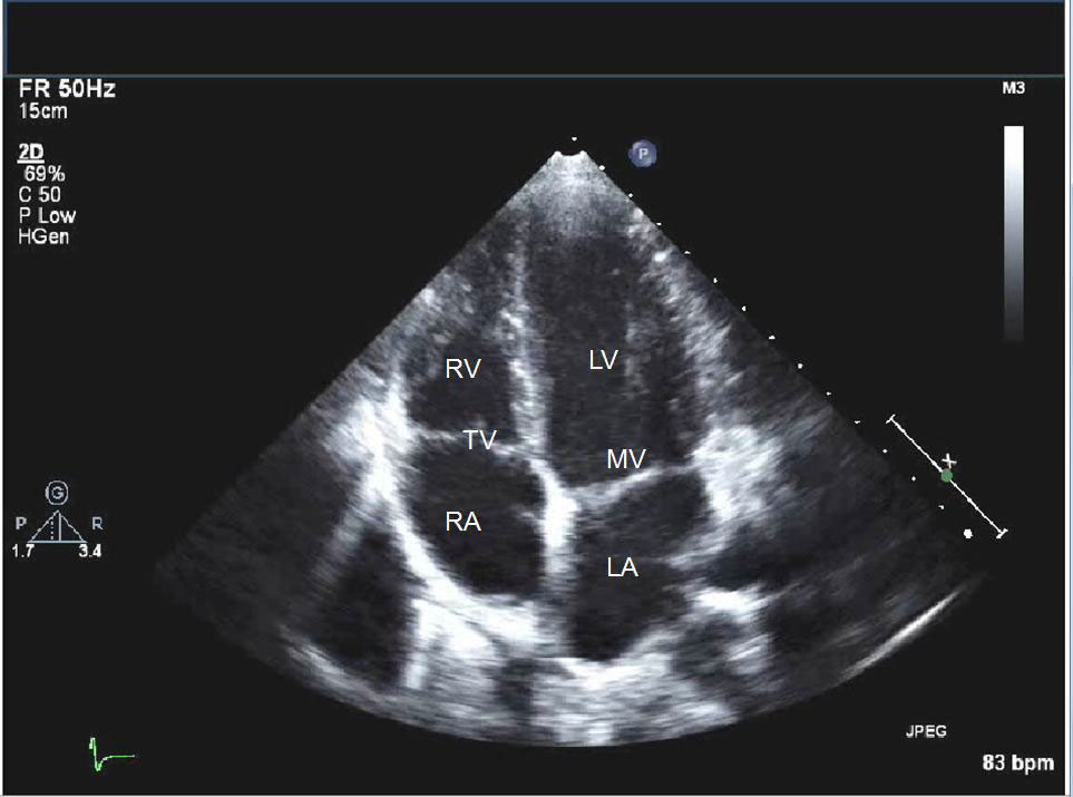
- RV - right ventricle
- TV - tricuspid valve
- RA - right atrium
- LV - left ventricle
- MV - mitral valve
- LA - left atrium
Computed Tomography can non-invasively diagnose coronary artery disease (CAD). The presence of calcified atheromatous plaques degrades evaluation for coronary luminal stenosis therefore this test is reserved for patients who have a low to intermediate pre test probability of CAD. Given its excellent spatial resolution, it is also a good test to assess the origin and course of the coronary arteries, in addition to luminal patency. It has a high negative predictive value and is therefore the test chosen to exclude CAD as the potential cause of symptoms.

Axial CT Angiogram image of a patient with history of prior myocardial infarction in the LAD territory.
Image demonstrates a peripherally calcified left ventricular apical aneurysm (BLUE arrows) with clot formation (RED arrow).
Myocardial Perfusion Testing is also an excellent test to evaluate for CAD but it cannot exclude the presence of atherosclerosis so it is reserved to assess flowlimiting disease in moderate to high-risk patients.

Magnetic Resonance Imaging is the gold standard test for evaluating the right ventricle. It is commonly used in patients with congenital heart diseases as it does not expose patients to ionizing radiation and better depicts the entire chest anatomy. These attributes make this modality ideal to follow these patients, particularly after surgical repair. The high tissue contrast resolution and ability to provide physiologic data makes this test unique when evaluating patients with cardiomyopathies and cardiac masses.


Cardiac catheterization is reserved for patients with acute myocardial infarction or severe coronary artery stenosis whom will likely require therapeutic intervention.
