Blood Smears Lab
Introduction
Blood provides a mechanism by which nutrients, gases, and wastes can be transported throughout the body. It consists of a number of cells suspended in a fluid medium known as plasma. Serum refers to plasma after clotting factors and fibrin have been removed.
Peripheral Blood Smear
The cells of the blood are important because they are a readily accessible population whose morphology, biochemistry, and ecology may give indications of a patient's general state or clues to the diagnosis of disease. For this reason, the complete blood count (CBC) and the white blood cell (WBC) differential are routinely used in clinical medicine. It is very important to be able to recognize normal blood cells and to distinguish pathological cells from the normal variants.
The identification of blood elements is based primarily on observations of the presence or absence of a nucleus and cytoplasmic granules. Other helpful features are cell size, nuclear size and shape, chromatin appearance, and cytoplasmic staining.
Component Cells of the Blood Smear
A blood smear is created by placing a drop of blood near the end of a clean glass microscope slide. Another slide is held at an angle, backed into the drop, and then smoothly dragged forward to spread the blood film along the slide. The blood must then be fixed, stained, and washed.
When you view a properly prepared blood smear of a healthy individual, there are several populations of cells that you will notice.
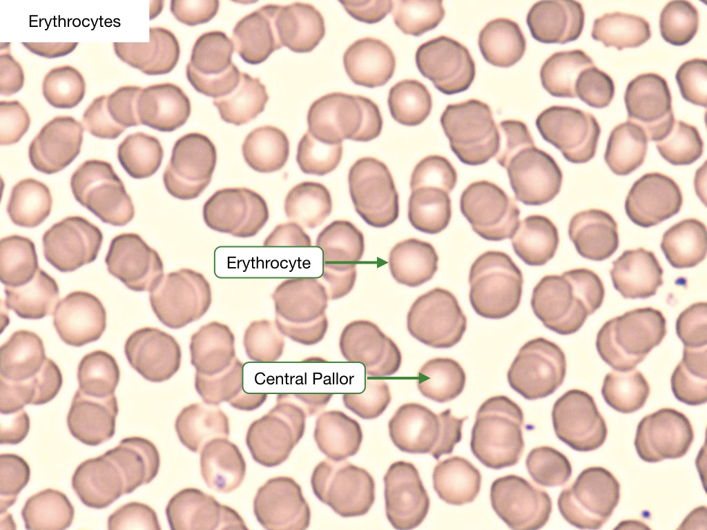
Erythrocytes
Erythrocytes, or red blood cells (RBC), are by far the predominant cell type in the blood smear. They are anucleate, non-granulated, eosinophilic cells that are uniform in shape (biconcave discs) and size (7.2 microns). RBCs have a central concavity that appears pale under the light microscope. These cells contain hemoglobin and are responsible for the transport and delivery of oxygen. RBCs have a lifespan of 120 days. The size of an RBC is measured automatically when a complete blood count (CBC) is performed; this is represented by the mean corpuscular volume (MCV), with normal being ~80-100 fL. Another useful RBC parameter measured on the CBC is red cell distribution width (RDW), which describes the size distribution of the entire RBC population, with normal being defined as less than 15%.
Reticulocytes
Reticulocytes are immature RBCs that are released from the bone marrow. They mature into RBCs after 1 to 2 days in the peripheral blood. There should be about one reticulocyte for every 100 red blood cells in a normal blood smear. These cells stain with a light blue tint because they still have RNA-containing organelles such as free ribosomes.
Thrombocytes
Thrombocytes, or platelets, are the smallest elements of the blood and are responsible for the formation of clots through a complex, highly regulated cascade. Platelets are between 2 and 5 microns in diameter and appear ovoid and anucleate with purple granules.

Leukocytes
Leukocytes, or WBCs, are cells of the immune system that are present in both blood and interstitial fluid. There should be about 1 leukocyte for every 1000 RBCs. They can be classified into two groups according to their nuclear pattern and the presence of cytoplasmic granules.
Monomorphonuclear leukocytes
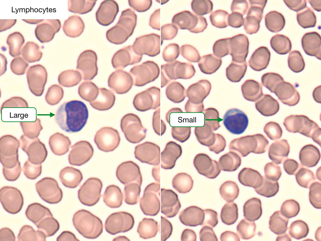
Monomorphonuclear leukocytes are cells with round, non-lobed nuclei. These include lymphocytes and monocytes.
Lymphocytes are about the same size as RBCs and have deeply stained nuclei with a thin rim of cytoplasm. The major population of lymphocytes is composed of B-cells and T-cells. A third population, known as natural killer (NK) cells, can also be identified. Lymphocyte counts are raised in response to viral infections.
Monocytes are larger than lymphocytes and have less-clearly demarcated nuclei that are usually not centered in the cell. These nuclei appear horseshoe-shaped, and the cytoplasm contains fine granules that give it a muddy gray color. These granules contain lysosomal enzyme and peroxidase. Monocytes are phagocytic cells that are important in the inflammatory response. They are the precursors to tissue macrophages.

Polymorphonuclear leukocytes
Polymorphonuclear leukocytes are cells with lobated nuclei and cytoplasmic granules. While these cells share the same primary (nonspecific) or azurophilic granules, they are named based upon the characteristics of their secondary (specific) granules.
Neutrophils are by far the most numerous of all WBCs. They are characterized by a nucleus that is segmented into three to five lobes that are joined by slender strands. The cytoplasm of neutrophils stains a pale pink. Its primary granules contain acid hydrolases and cationic proteins, and its secondary granules contain a variety of antimicrobial substances used to destroy bacteria that they phagocytose during the acute inflammatory response.

Eosinophils are larger than neutrophils and are distinguished by large red or orange granules of uniform size. These granules contain major basic protein, which is released to kill organisms too large to phagocytose, such as parasites and helminths.

Basophils are intermediate in size between neutrophils and eosinophils and have simple or bilobed nuclei. They contain many coarse purple granules that can vary in size or shape. These granules contain histamine, which is released to cause a vasoactive response in hypersensitivity reactions, and heparin, which is an anticoagulant. Basophils are not phagocytic.
